Arterial Pressure Waveforms
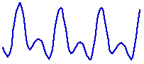
The pumping of the heart results in the development of pressure in the aorta and the arteries. If pressure in the aorta is recorded over time a pressure wave can be observed:

Many factors influence the aortic pressure waveform. Consider the following example:

Greater ventricular filling (more filling time) resulted in greater systolic pressure at C compared with E. This is explained by the Frank/Starling mechanism. Starling stated that "the energy of contraction is a function of the length of the muscle fibre." So the greater the filling of the ventricles the stronger the subsequent systolic contraction.
Notice that the diastolic pressure at B is lower than the diastolic pressure at D. This is a function of the time available for blood to flow out of the aorta and for the pressure in the aorta to fall.
Other factors such as aortic valve conditions, compliance (elasticity) of the aorta (related to age and disease), vascular resistance, cardiac output and technical considerations of recording can affect the arterial pressure waveform.
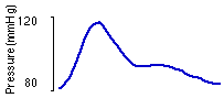
As pressure waves travel from the aorta and large arteries to the narrower, less compliant distal arteries they travel at a greater speed. The ascending part of the wave (anacrotic limb) becomes steeper and the maximum systolic pressure tends to increase.
The pulse pressure wave on the left was recorded in a smaller peripheral artery, while the pressure wave on the right was recorded from the aorta.
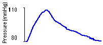
Notice the dip in the arterial pressure waveform occurring on the descending part of the wave (dicrotic limb). This is referred to as the dicrotic notch. The dicrotic notch in an arterial pressure waveform does not necessarily correspond to the incisura in the aortic pressure waveform (caused by closure of the aortic valve). The dicrotic notch and the dicrotic wave that follow it are thought to be due to a reflected pressure wave. The depth of the dicrotic notch appears to increase following infusion of vasodilators, as demonstrated by the below waveform that was recorded after infusion of hydralazine.