Abnormal EKG’s and Corresponding Arterial Waveforms
Tachycardia
Supraventricular
Paroxysmal Tachycardia
Ventricular Fibrillation
The Normal Sinus Rhythm
Normal sinus rhythm refers to the usual case in healthy adults where the SA node is the cardiac pacemaker and the heart rate is 60 - 100 beats per minute (BPM).
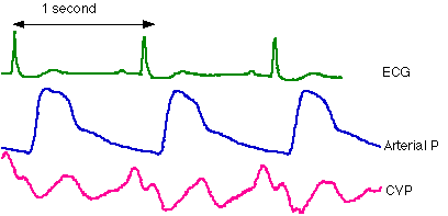
With a normal sinus rhythm one would expect
the following:
- a normal ECG with P, QRS and T waves
- a PR interval of 0.12 - 0.20 seconds
- a regular interval between each QRS complex (R-R interval) of 0.60 - 1.00 seconds
- a distinct a wave in the CVP trace due to atrial contraction
- a regular arterial pulse pressure (difference between systolic and diastolic pressures)
- there may be minor regular variation in heart rate and pulse pressure associated with respiration (sinus arrhythmia)
However it is often the case in the clinical
situation that the patient does not have a normal sinus rhythm and it is
important to be able to recognize and identify these abnormal cases. The
following pages of this tutorial will describe and illustrate various abnormal
situations.
Sinus Bradycardia
Sinus bradycardia refers to a sinus rhythm
(ie originating in the SA node) which is slower than 60 BPM. (brady -
slow, cardia - heart). Sinus bradycardia may occur with vagal
(parasympathetic) stimulation, such as in trained athletes or in patients with
the carotid sinus syndrome (in whom baroreceptors are overly sensitive to
pressure, resulting in excessive vagal stimulation). Sinus bradycardia may also
occur as a result of pharmacological beta-blockade.
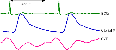
The rhythm is similar to normal sinus rhythm,
except that the R-R interval is greater than one second. The pulse pressure may
be greater due to a greater stroke volume (resulting in greater systolic
pressure) and increased time for diastolic run-off (resulting in lower
diastolic pressure).
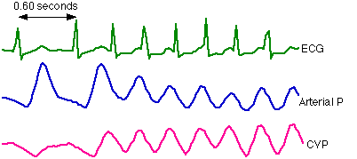
Sinus Tachycardia
Sinus tachycardia refers to a sinus rhythm
with a heart rate greater than 100 BPM (tachy - fast). Sinus tachycardia
may be due to fever, which results in increased excitability of the SA node.
Sympathetic stimulation (from a variety of causes) and cardiac toxicity may
also cause sinus tachycardia.

The rhythm is similar to normal sinus rhythm,
except that the R-R interval is less than 0.6 seconds. The a wave may
tend to merge with the v wave in the CVP trace, and the P wave may be
obscured by the T wave in the ECG (not apparent in this trace). The pulse
pressure may be lesser due to a lower stroke volume and decreased time for
diastolic run-off.
Atrial Fibrillation
In atrial fibrillation small areas of atrial
tissue repeatedly depolarize but in a disordered way relative to neighboring
areas of atrial tissue. This is believed to involve a microreentry mechanism.
There is no concerted depolarization or contraction of the atria. Also, due to
the chaotic nature of atrial depolarizations, there is irregular penetration of
the AV node, resulting in irregular ventricular contractions.
Atrial fibrillation is most common in individuals with atrial enlargement, often associated with valvular pathologies.
Notice the lack of P waves due to continual,
irregular depolarisations of different areas of atrial tissue. The QRS
complexes have normal shape, due to normal ventricular conduction, however the
R-R intervals vary from beat to beat with no regular pattern.
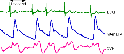
Notice that the systolic arterial pressure
varies from beat to beat as ventricular filling time changes. Also the
diastolic pressure changes from beat to beat with changes in diastolic runoff
time. So the pulse pressure also may vary significantly from beat to beat.
Key features of atrial fibrillation:
- absence of P waves
- irregular R-R interval
- varying pulse pressure
- no a waves in the CVP trace
Ventricular Ectopics
ectopic
- 'in abnormal place or position' from the Greek 'ektopos' - out of place.
An ectopic beat occurs from an abnormal site
(called an ectopic focus) before the expected time of the next contraction.
Ectopic beats (also called extrasystoles or premature contractions) may
originate in the atria, the AV junction or the ventricles. There are numerous
possible causes of ectopics including local ischaemia, drugs (caffeine is a
good example), calcified plaques and physical contact (such as contact of the
heart with catheters or surgical instruments).
Take a close look at the following diagram:

This ventricular ectopic - or premature
ventricular contraction (PVC) - has had an effect on all three traces in the
above diagram:
The ECG
- QRS earlier than expected (premature) i.e. shorter RR interval than normal
- QRS wider than normal
- QRS voltage higher than normal
- inverted T wave
- obscured P wave
- next RR interval longer than normal
The arterial pressure
- ejection earlier than expected
- low systolic pressure generated
The CVP
- large a wave
- a wave at expected time (not premature)
These observations can be explained by
considering the abnormal pathway of ventricular depolarization:
The depolarization of the ventricles was
initiated prematurely at an ectopic focus, not a part of the His-Purkinje
network, so the wave of depolarization traveled slower through the myocardium
via unconventional pathways, resulting in a wide, unusually-shaped QRS.
The slow conduction resulted in a less
concerted contraction of the ventricles. This, as well as less time for
ventricular filling and lack of atrial priming of the ventricles (ventricles
depolarized before the atria), accounts for the poor ejection and low systolic
arterial pressure.
When the ventricles depolarize normally,
voltages on one side of the heart tend to be balanced by voltages on the other
(due to the heart's symmetry). However with an ectopic depolarization voltages
on one side may have no counterpart, due to the slower propagation of the wave
of depolarization, resulting in a greater measured voltage in the ECG. That is
why a PVC can result in a QRS with a higher voltage than normal.
The slow ventricular depolarization resulted
in those areas of myocardium first depolarised being able to repolarize first
(in contrast with the case of a normal depolarization). This pattern of
repolarization resulted in the inverted T wave.
The SA node fired at the expected time. The
atria contracted but, because the ventricles were already contracting, they
were unable to eject into the ventricles, resulting in markedly increased
pressure in the atria and hence a large CVP a wave (cannon wave). The a
wave was larger than normal but occurred at the expected time.
The two waves of depolarization (one from the
SA node and the other from the ectopic focus) met in the AV node. The ectopic
depolarization did not reset the SA node, so the next SA node firing occurred
exactly as if there had been no ectopic depolarization. The next RR interval,
by being longer than normal, exactly compensated for the prematurity of the
ectopic . This is termed a fully compensatory pause.
Characteristics of (most) PVCs
- wide and bizarre QRS, often with a high voltage and inverted T wave
- reduced or no left ventricular ejection
- large CVP a wave
- fully compensatory pause
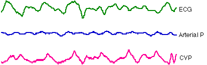
Supraventricular Paroxysmal Tachycardia
Take a close look at the following diagram:
Note the sudden onset of tachycardia which
results in a decreased mean arterial pressure (due to decreased filling time)
Also note that the QRS complexes are not
abnormal in shape, indicating a supraventricular irritable focus.
Ventricular Fibrillation
Ventricular fibrillation is analogous to
atrial fibrillation and similar phenomena are believed to give rise to it.
However ventricular fibrillation is a very serious condition because the
uncoordinated contractions of ventricular myocardium result in ineffective pumping.
If immediate action is not taken the results are fatal.
These traces were recorded in a patient
undergoing cardiopulmonary bypass, so that blood flow was maintained
mechanically.
The following features can be seen in the
above diagram :
- random, unrelated waves in the ECG
- lack of significant pulse
pressure